
Technology development in service of patients
Four programs, each pairing a measurement we build with a question that could not be answered without it. Most of the questions come from the clinic, where a small number of cells that survive treatment, or that quietly become cancer, decide a patient's outcome.
More from a single cell, and still adding layers
Every program below depends on measurements that did not exist a few years ago. This program builds them, and each of the others is an application of it.
The lab invented primary template-directed amplification (PTA), which amplifies the genome of a single cell far more uniformly than earlier methods and lets us call roughly 90 percent of small variants in a cell with high precision. We then paired PTA with full-length transcript capture so that genomic variants and transcript abundance are read from the same cell, and we are now adding DNA methylation, through a 5-methylcytosine-selective deaminase applied before amplification, and surface proteins. No method reads everything in a cell. Targeted panels return the driver genotype and a few hundred transcripts, while our methods return the genome, the transcriptome, and now the methylation of that same cell, and we are still adding layers, at a cost that allows thousands of cells per patient. In parallel, we build wet-lab and computational methods to detect mutations, methylation changes, and microbial DNA in cell-free DNA from blood.
This is where a chemist, an engineer, or a computational biologist joining the lab makes the largest difference. A new chemistry here changes what every other program can see.
Where we are: PTA and same-cell genome plus transcriptome sequencing are in use by laboratories worldwide and are available commercially as ResolveDNA and ResolveOME; the methylation readout is in development.
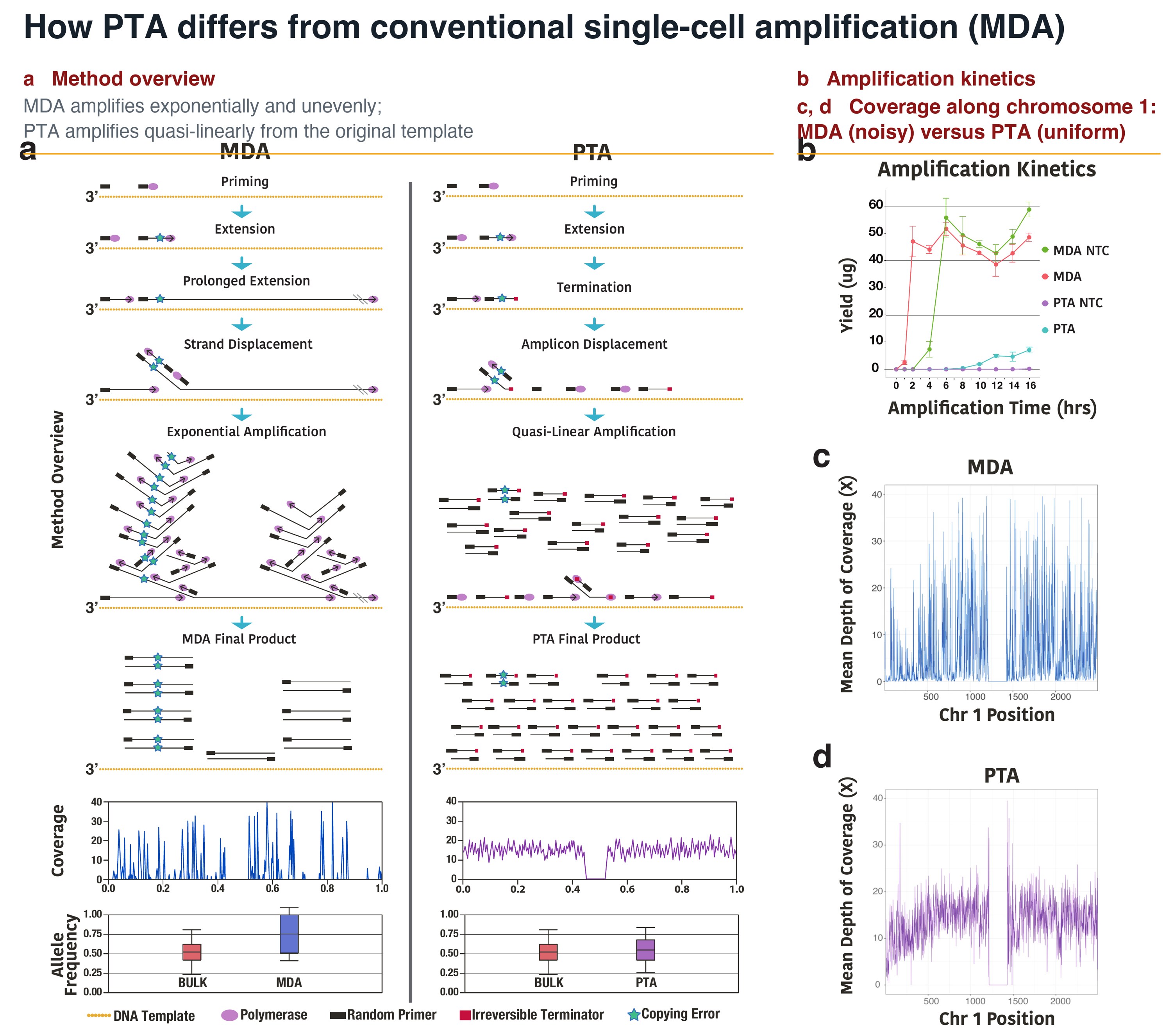
How PTA works. In MDA, exponential amplification from the first primed sites over-represents random loci; in PTA, exonuclease-resistant terminators keep amplification quasi-linear from the original template, giving far more uniform coverage. From Gonzalez-Pena et al., PNAS 2021.
Outrunning relapse in childhood leukemia
Most children with acute lymphoblastic leukemia are cured, but 10 to 15 percent are not, and only about half of the children who relapse survive. Bulk sequencing of their leukemia finds little to explain why, because the cells that survive months of chemotherapy are a tiny minority hidden among millions.
We sequence those cells one at a time. With PTA we found that individual leukemia cells carry 3.7 times more mutations than bulk sequencing detects, that patients thought to have a single RAS mutation carry a median of five in separate clones, and that the clones which persist after induction therapy can be traced by their genomes and surface markers. We are now testing whether noncoding mutations in regulatory DNA change the state of individual cells in ways that let them survive treatment, in a cohort of children with measurable residual disease, and validating candidate mutations with pooled CRISPR screens. The same approach is being extended to acute myeloid leukemia.
The goal is a single-cell test that flags relapse months before it is clinically visible, and a catalog of resistance mutations that guides each child to the most effective, least toxic therapy.
Where we are: patient samples come from clinical trials at St. Jude Children's Research Hospital and Stanford; collaborators Dan Landau (Weill Cornell) and William Evans (St. Jude).
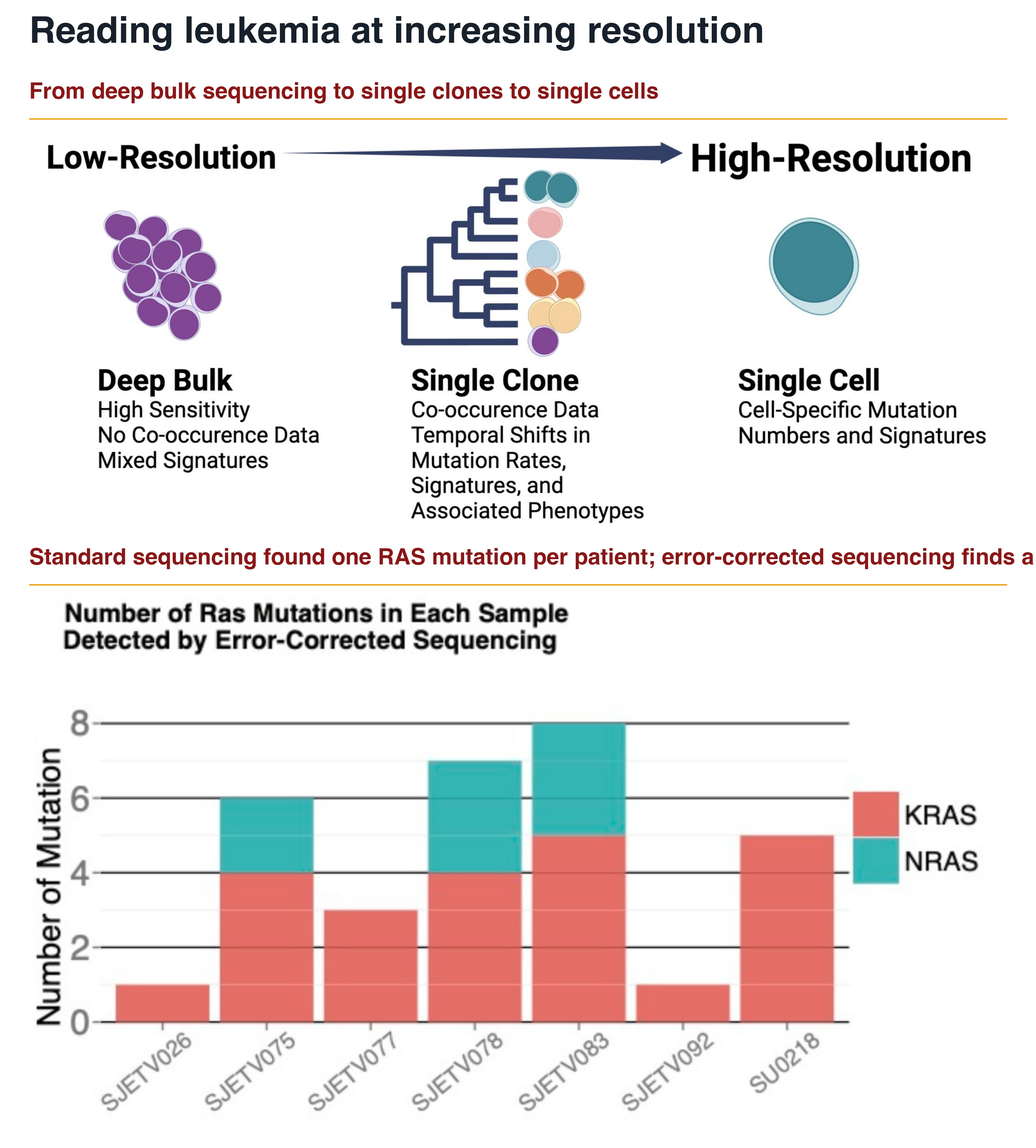
Why single cells matter. Bulk sequencing sees one RAS mutation per patient; error-corrected and single-cell sequencing reveal a median of five, and individual leukemia cells carry 3.7 times more mutations than bulk sequencing detects. From Pang, Prieto et al., bioRxiv 2025 (v2).
Do common childhood viruses cause leukemia?
The most common initiating lesion in childhood acute lymphoblastic leukemia, the ETV6-RUNX1 fusion, arises before birth in about one in a hundred newborns, yet only about one in a hundred of those children ever develops leukemia. Something after birth supplies the second hits. Mel Greaves' delayed-infection hypothesis has long pointed at childhood infections, and mouse models confirm that pathogen exposure is required, but the mechanism by which an infection damages the genome of a human preleukemic cell has remained unknown.
The genomes of these leukemias carry two tell-tale scars: point mutations with the signature of the antiviral enzyme APOBEC3A, and deletions with the signature of RAG, the enzyme that rearranges antibody genes. We are testing whether common respiratory viruses such as influenza infect B-cell precursors directly and, through interferon signaling, switch on APOBEC3A while RAG is active, producing exactly the mutations seen in patients. Using cord-blood precursors carrying ETV6-RUNX1, staged B-cell differentiation, and same-cell genome and transcriptome sequencing, we can read viral infection, interferon response, mutational signatures, and deletions in the same single cells.
If the mechanism holds, it points toward prevention. The infections that deliver the second hit are ones medicine already knows how to reduce.
Where we are: an ex vivo model of viral leukemogenesis is running in the lab, with primary patient samples from St. Jude Children's Research Hospital.
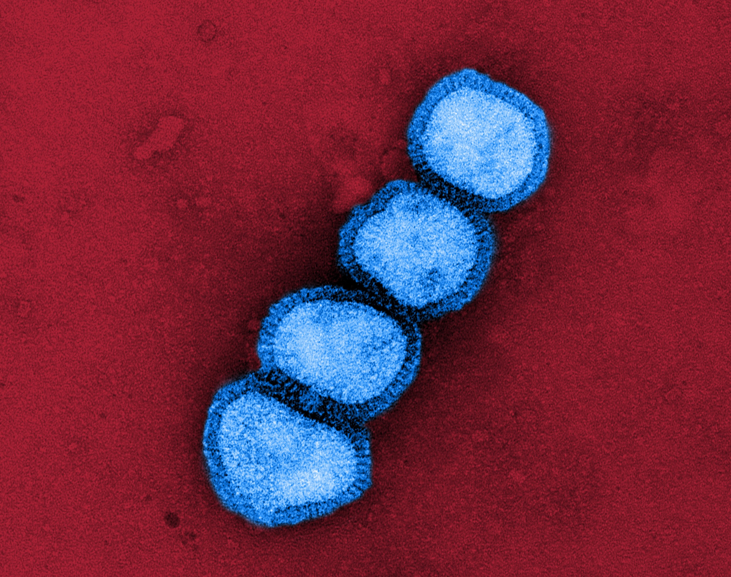
Influenza A (H1N1) virus particles, colorized transmission electron micrograph. Image: NIAID, public domain.
When a disease lives in only some of a child's cells
Every one of us is a mosaic. From the first cell division onward, each cell acquires its own mutations, so no two cells in the body carry exactly the same genome. Usually that is harmless. But when a mutation arises early in development and lands in the wrong cells, it can cause epilepsy, overgrowth of one part of the body, vascular or lymphatic malformations, or other conditions that standard genetic testing can miss, because the mutation is absent from the blood sample the test was run on.
We built our single-cell methods for exactly this problem. By sequencing the genomes of individual cells from the affected tissue, we can find a mutation that is present in only some cells, learn which tissues carry it and when in development it arose, and give a family an answer that a normal blood test could not. In a child with severe epilepsy, we sequenced single neuronal nuclei recovered from the tips of stereo-EEG electrodes placed for surgical planning, to look for mutations confined to the part of the brain that was seizing.
The same mosaicism, accumulated over a lifetime, is where cancer begins. We use the same tools on normal and precancerous tissue to date when a clone arose, measure how quickly it gathers genetic and epigenetic damage, and ask what the immune system does about it, with the aim of catching cancer before it starts. A related effort measures directly, in human blood stem cells and without animals, how much genomic damage a chemical exposure causes.
Collaborators: Gerald Grant (Duke Neurosurgery), Christopher Walsh and Peter Park (Harvard).
Stereo-EEG electrodes mapped onto a pediatric patient's brain. After the electrodes are removed, we sequence the genome of single neuronal nuclei from their tips. Collaboration with Gerald Grant, Duke Neurosurgery.
Where the tools are going next
Smaller projects that apply the same single-cell chemistry to new problems. Each is a good entry point for a rotation student or a new collaborator.
Single-microbe genomics
PTA adapted to single bacterial cells recovers near-complete genomes without culture, in collaboration with the DOE Joint Genome Institute. We are now pointing it at the microbes that live inside tumors and at antibiotic resistance in the airways of children with cystic fibrosis, with Christin Kuo at Stanford.
Circulating tumor cells
Same-cell genome, transcriptome, and surface protein from the rare tumor cells captured in a blood draw, to follow how a cancer spreads and evades the immune system, with Stanford's Center for Cancer Systems Biology.
Infection diagnostics from cell-free DNA
Sequencing microbial DNA in plasma identified bloodstream infections in children with leukemia days before symptoms appeared, with St. Jude Children's Research Hospital.
Methods that leave the building
We build technologies so that they can be used, and we measure success partly by how far they travel. PTA is now a commercial product used by academic and industry groups worldwide, it is licensed for preimplantation genetic testing in IVF clinics, and paired single-cell genome and transcriptome sequencing of residual leukemia cells, an approach developed here, is offered as a service to other researchers. Chuck Gawad co-founded BioSkryb Genomics to carry that work out of the lab and serves as its scientific founder.
Interested in collaborating?
We are always excited to work with new scientists. Email Chuck Gawad with a short description of the question you want to answer.